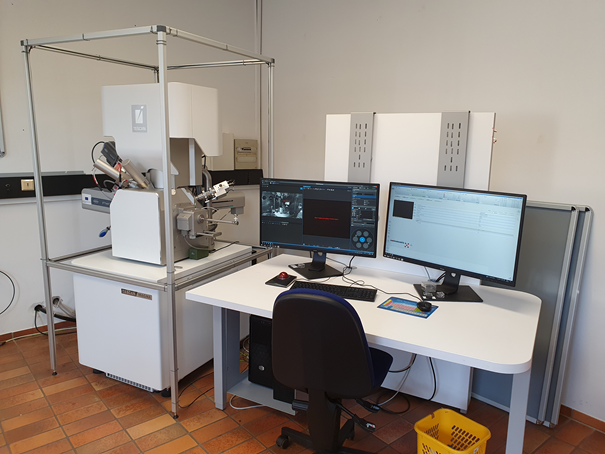
SEM Tescan MAGNA
The scanning electron microscope MAGNA from Tescan is a powerful tool for imaging in ultrahigh resolution. With each immersion optics and crossover-free electron beam it allows excellent imaging, also at low electron energies. With its manifold detectors
- Everhart-Thornley secondary electron detector
- In column secondary electron detector
- 4 quadrant backscatter detector
- Low-energy backscatter detector
- Scanning transmission detector, subdivided into four ring segments
it allows investigation of all kind of materials, investigated at the institute. The beam deceleration option and a “low vacuum modus” (chamber pressure up to 500 Pa) widen the imaging possibilities of this microscope.
- Electron backscatter diffraction detector (Bruker)
- Electron dispersive X-ray detector (Bruker)
are also attached to the SEM, complementing qualitative microstructural investigation capabilities with quantitative access onto chemistry and microstructure.
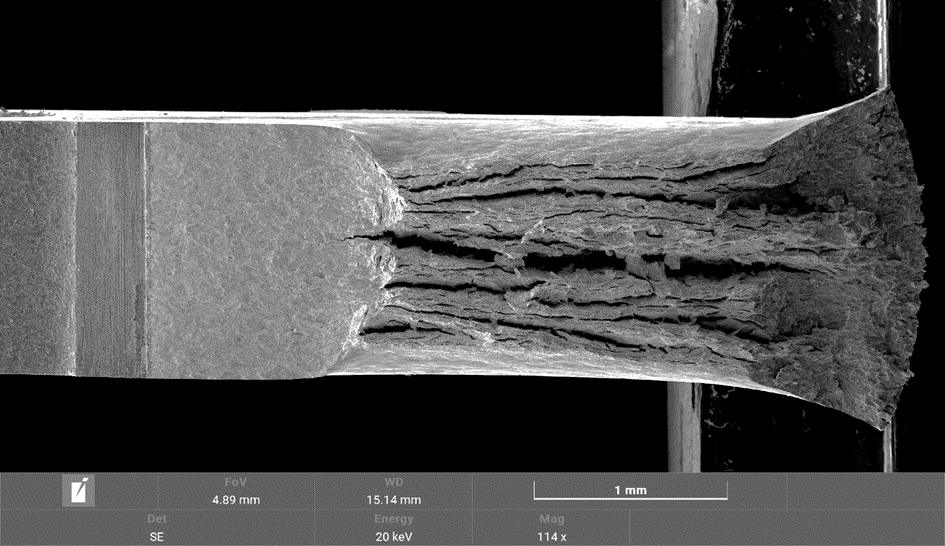
Fracture Surface of W sample, after a fatigue crack growth experiment

Back scatter detector micrograph of the microstructure of Ta, severe plastically deformed using high-pressure torsion www.oeaw.ac.at/esi/research/nanomaterials-by-severe-plastic-deformation

Dark-field STEM image of Ti, severe plastically deformed using HPT